
Technical support for imaging and analysis
The Center for Quantitative Imaging provides advanced X-ray computed tomography and image analysis for researchers, industry partners and government collaborators.
This page organizes the center's capabilities around what users need to understand: what can be imaged, which systems and techniques are available, what sample types can be submitted and what kinds of data and visualizations partners receive.

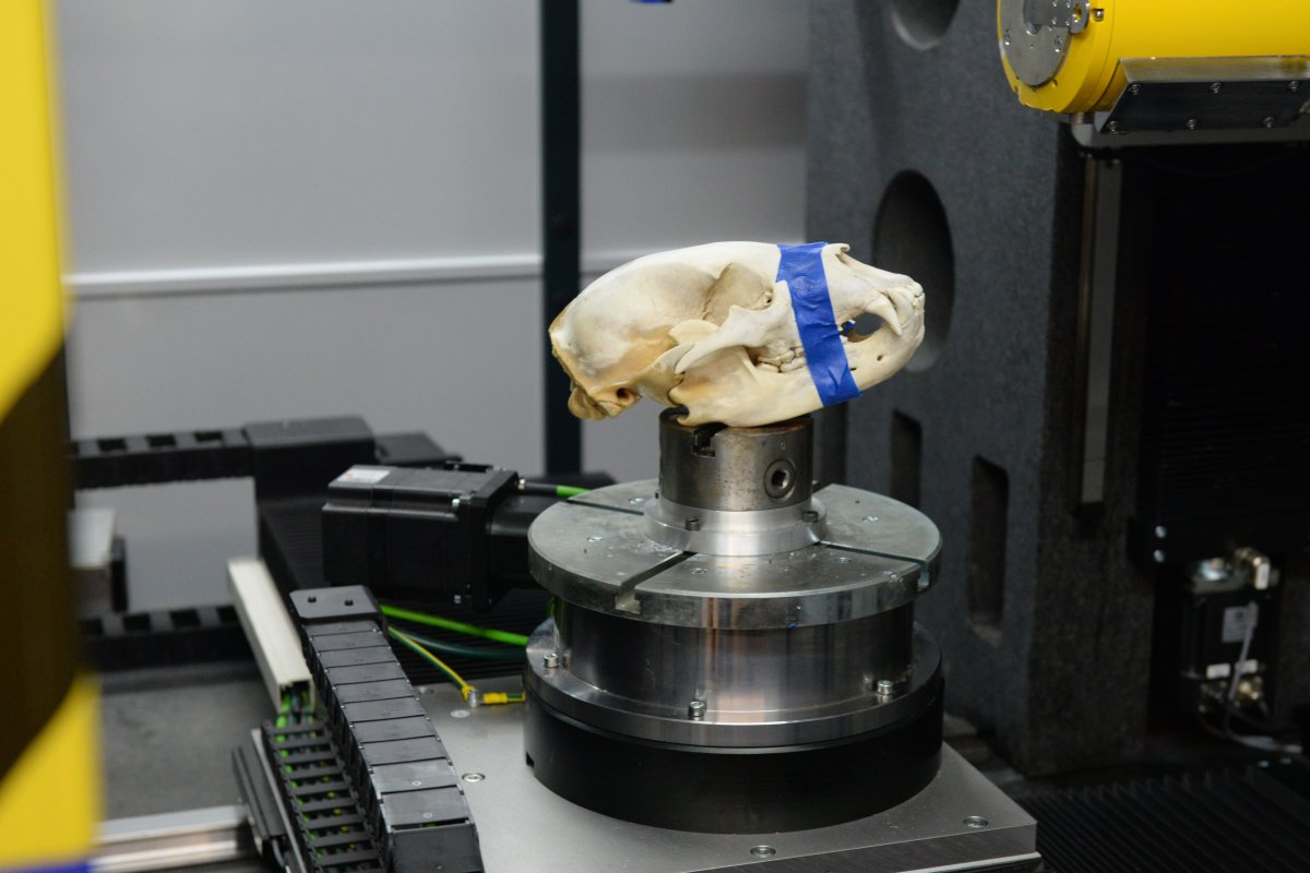
Capabilities
CT scanning, visualization and measurement.
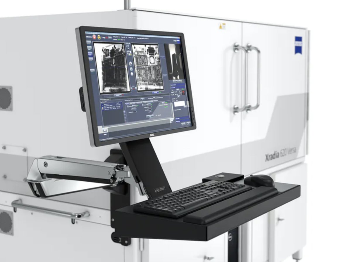
Equipment
Systems used to support scanning and analysis.

Techniques
How project questions connect to imaging workflows.

Sample types
Materials, biological specimens and products.

Deliverables
Datasets, reconstructions and measurements.
Capabilities

3D Visualization and Analysis
Processing and analysis of CT images to extract quantitative and qualitative sample information.
- Porosity and fracture measurements
- Particle size and material distribution
- TIFF, STL, and VOL data outputs
- 3D video rendering and virtual tours
- Advanced image processing and visualization

X-ray computed tomography
Non-destructive imaging that reveals internal structures without cutting, sectioning or otherwise altering samples.
- High-resolution 2D and 3D imaging
- Internal structure visualization
- Non-destructive evaluation
- Comparative scans and repeat imaging
Quantitative image analysis
Image processing and measurement workflows that turn CT datasets into usable research and engineering data.
- Segmentation and feature extraction
- Volume and dimensional measurements
- Porosity and void analysis
- Defect and inclusion characterization

Research collaboration
Staff expertise helps connect imaging questions to the right scan strategy, analysis workflow and deliverable.
- Project planning and feasibility review
- Sample preparation guidance
- Method development
- Data interpretation support
Equipment
Zeiss Versa 620
Non-destructive sub-micron scale imaging for intact samples and advanced materials.
- True spatial resolution of 500 nm
- Minimum achievable voxel size of 40 nm
- In-situ tensile and compression testing support
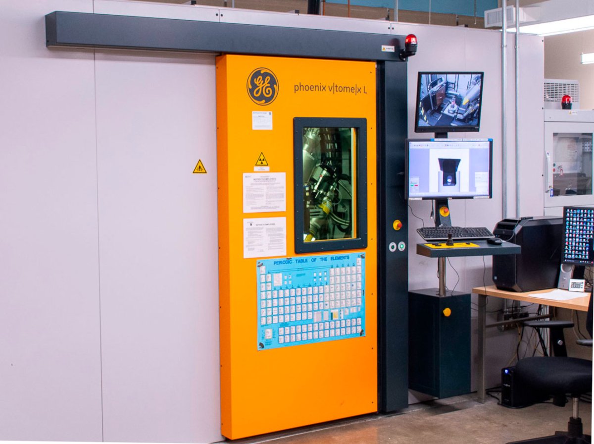
GE v|tome|x L Micro-Nano CT
High-resolution, 3D-computed tomography for a range of object sizes and densities.
- Dual X-ray tube system
- High-energy scanning for dense objects
- Nanofocus imaging for sub-micron detail
Techniques and Workflows
Imaging workflows are selected based on the research question, sample size, material density, resolution needs and expected deliverables.
This section organizes techniques by what partners are trying to accomplish rather than by equipment alone.

Non-destructive internal imaging
Used when a project requires internal visualization of structures, defects, voids, inclusions or assemblies without damaging the sample.
3D reconstruction and visualization
Used to generate digital volumes, rendered views and visual outputs that help researchers inspect and communicate internal structure.
Segmentation and measurement
Used to isolate features of interest and quantify volume, porosity, dimensions, density variation or spatial relationships.
Custom imaging workflows
Used when unusual sample types, experimental designs or analysis goals require tailored imaging and processing methods.
Common Sample Types
Materials and products
- Polymers and composites
- Manufactured parts
- Coatings and assemblies
- Packaging and engineered materials
Biological specimens
- Plant structures
- Animal tissues
- Bone and hard tissue
- Small biological samples
Earth and environmental samples
- Soils and cores
- Rocks and minerals
- Porous media
- Environmental materials
Research and prototype samples
- Experimental materials
- Prototype components
- Multi-material assemblies
- Custom research samples
Data, Analysis, and Deliverables
What partners receive
Project deliverables depend on the scan type, sample properties and project goals.
- CT datasets and reconstructed image stacks
- 2D slices and 3D renderings
- Segmented features or regions of interest
- Quantitative measurements and analysis outputs
How staff support projects
Staff help connect research questions to appropriate imaging workflows.
- Review project fit and feasibility
- Discuss sample size, density and preparation needs
- Support scan planning and analysis workflows
- Coordinate reporting expectations and next steps
Ready to discuss a project?
Work with staff to determine the right imaging approach, sample requirements and next steps for your research or partner project.