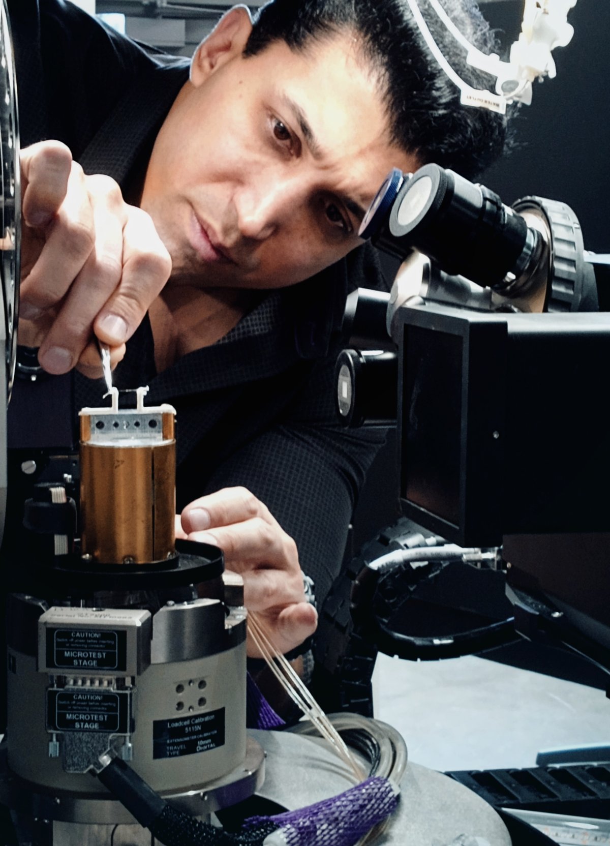
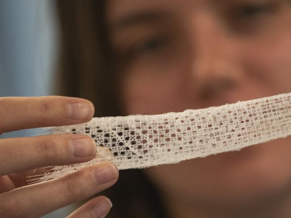
Fariborz Tavangarian, associate professor of mechanical engineering at Penn State Harrisburg, is investigating the intricate structure of a marine sponge (Eulectella aspergillum) to develop innovative materials for human bone tissue engineering. Despite being composed of fragile silica, the sponge exhibits remarkable strength due to its unique layered architecture. To unravel the secrets of this natural wonder, Tavangarian is collaborating with the Center for Quantitative Imaging's Michelle Quigley and Andrew Ross to analyze the sponge's individual fibers, known as spicules. By analyzing how the spicules withstand stress and break, the researcher aims to inform the creation of bio-inspired materials with superior strength and durability for biomedical applications.

“The Center for Quantitative Imaging enables us to test the sponge fibers and directly scan them without any post-processing or modification. This capability is essential to our research, as post-processing steps on fractured spicules could alter the findings. CT scanning allows us to observe the results in their original, unaltered state.”
— Fariborz Tavangarian, associate professor of mechanical engineering at Penn State Harrisburg


